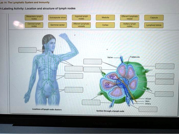
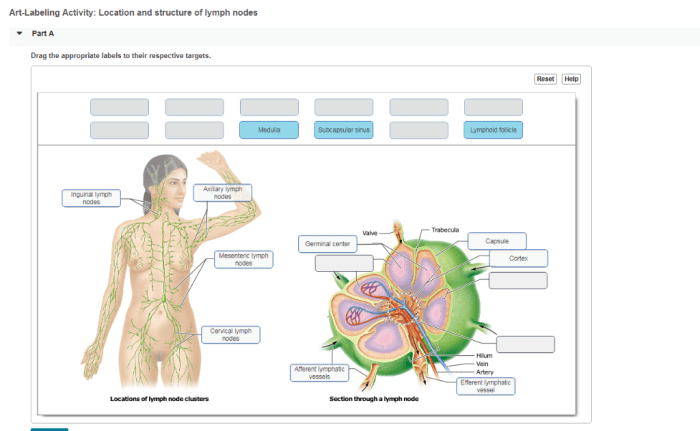
Art-labeling activity: location and structure of lymph nodes – Embark on an art-labeling activity that unravels the intricate world of lymph nodes, their strategic positioning within our bodies, and their crucial role in our immune system. This exploration promises to illuminate the fascinating structure of these immunological guardians, shedding light on their functions and clinical significance.
Lymph nodes, scattered throughout our bodies like vigilant sentries, play a pivotal role in filtering and monitoring bodily fluids. Their strategic placement near major blood vessels, organs, and body cavities highlights their importance in immune surveillance. This activity delves into the regional classification of lymph nodes, distinguishing between superficial and deep nodes, and explores the histological organization of these nodes, including the capsule, cortex, paracortex, medulla, and hilum.
Location of Lymph Nodes

Lymph nodes are small, bean-shaped structures that are distributed throughout the body and play a crucial role in the immune system. They are strategically positioned in close proximity to major blood vessels, organs, and body cavities, allowing them to efficiently filter and monitor the surrounding tissues and fluids.
Regional Classification of Lymph Nodes
Lymph nodes can be classified into two main categories based on their location:
- Superficial lymph nodes: Located just beneath the skin and are easily palpable, such as the axillary (armpit) and inguinal (groin) nodes.
- Deep lymph nodes: Located deep within the body, often associated with specific organs or structures, such as the mediastinal (chest) and intra-abdominal (within the abdomen) nodes.
Structure of Lymph Nodes

Lymph nodes have a highly organized histological structure that facilitates their immune functions:
- Capsule: A tough, fibrous outer layer that surrounds and protects the lymph node.
- Cortex: The outermost layer, containing densely packed lymphocytes, including B cells and T cells, which are responsible for recognizing and responding to foreign antigens.
- Paracortex: A transitional zone between the cortex and medulla, containing specialized cells called dendritic cells that present antigens to lymphocytes.
- Medulla: The innermost layer, containing sinuses where lymphocytes and macrophages filter and remove foreign particles and pathogens.
- Hilum: A depression on one side of the lymph node where blood vessels and lymphatic vessels enter and exit.
Interconnections and Drainage Patterns: Art-labeling Activity: Location And Structure Of Lymph Nodes

Lymph nodes are interconnected by a complex network of lymphatic vessels that transport lymph, a fluid containing immune cells and waste products, throughout the body.
Lymph flows in a unidirectional manner, from the tissues into the lymphatic vessels and then through the lymph nodes, where it is filtered and immune responses are initiated. The direction of lymph flow determines the drainage pattern, which varies for different regions of the body.
Clinical Significance of Drainage Patterns
Understanding lymphatic drainage patterns is crucial in clinical practice, as it helps guide surgical procedures and predict the spread of infections or malignancies. For example, surgeons use sentinel lymph node mapping to identify the first lymph node(s) that receive lymphatic drainage from a tumor, which can aid in staging and treatment planning.
Clinical Applications
Lymph nodes play a vital role in physical diagnosis and surgical oncology:
Lymph Node Examination, Art-labeling activity: location and structure of lymph nodes
Palpation of lymph nodes during a physical examination can provide valuable information about their size, consistency, and tenderness, which can help diagnose infections, malignancies, and other pathological conditions.
Lymph Node Biopsy
Lymph node biopsy involves removing a small sample of tissue for microscopic examination. This procedure is used to confirm a diagnosis of infection or malignancy and to assess the extent of disease.
Lymph Node Mapping and Sentinel Lymph Node Identification
Lymph node mapping and sentinel lymph node identification are techniques used in surgical oncology to determine the extent of tumor spread and guide surgical treatment. Sentinel lymph nodes are the first lymph nodes that receive lymphatic drainage from a tumor, and their status can provide prognostic information and guide surgical decisions.
Helpful Answers
What is the function of lymph nodes?
Lymph nodes act as filters, trapping and removing foreign substances, pathogens, and cellular debris from the lymphatic fluid.
How are lymph nodes classified?
Lymph nodes are classified based on their location, either superficial (located just beneath the skin) or deep (located within body cavities).
What is the significance of lymph node mapping?
Lymph node mapping helps identify the sentinel lymph node, which is the first node to receive lymphatic drainage from a particular region. This is important in surgical oncology, as it guides the extent of lymph node removal during cancer surgery.