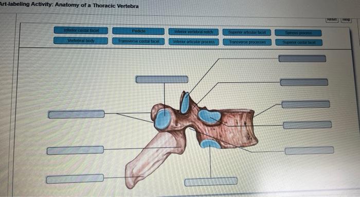
Art-labeling activity anatomy of a thoracic vertebra – Embark on an immersive journey into the intricate anatomy of a thoracic vertebra, guided by our comprehensive art-labeling activity. As we delve into the intricacies of this spinal building block, prepare to uncover its remarkable structure, function, and clinical significance.
This activity is designed to provide a comprehensive understanding of the thoracic vertebra, its anatomical landmarks, and its role within the spinal column. Through detailed descriptions and interactive labeling exercises, you will gain a deeper appreciation for the complexities of human anatomy.
Anatomical Landmarks

Thoracic vertebrae are located in the middle section of the spinal column, between the cervical vertebrae above and the lumbar vertebrae below. They are responsible for providing support and protection to the thoracic cavity, which houses the heart and lungs.
Vertebral Body
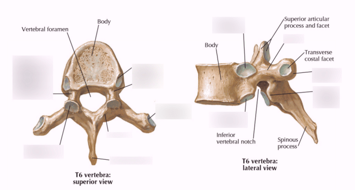
The vertebral body is the main weight-bearing component of the thoracic vertebra. It is shaped like a cylinder with a slightly concave anterior surface and a convex posterior surface. The superior and inferior surfaces of the vertebral body are flattened and covered with cartilage, which allows for articulation with adjacent vertebrae.
The vertebral body plays a crucial role in weight-bearing and shock absorption. It is made of dense bone that can withstand significant compressive forces. The superior and inferior vertebral notches provide attachment points for the intervertebral discs, which further enhance shock absorption and provide flexibility to the spinal column.
Spinous Process
The spinous process is a bony projection that extends posteriorly from the vertebral arch. It is shaped like a triangle with a broad base and a pointed tip. The spinous process serves as a lever arm for muscle attachment, providing support and stability to the spinal column.
The length and shape of the spinous process vary among different thoracic vertebrae. The spinous processes of the upper thoracic vertebrae are longer and more pointed, while those of the lower thoracic vertebrae are shorter and more blunt.
Transverse Processes, Art-labeling activity anatomy of a thoracic vertebra
The transverse processes are two bony projections that extend laterally from the vertebral arch. They are shaped like flattened ovals with a concave anterior surface and a convex posterior surface. The transverse processes provide attachment points for the ribs.
Together with the pedicles and laminae, the transverse processes form the intervertebral foramina, which are openings through which the spinal nerves and blood vessels pass.
Articular Processes
The articular processes are four bony projections that extend from the vertebral arch. The superior articular processes are located on the superior aspect of the vertebral arch, while the inferior articular processes are located on the inferior aspect of the vertebral arch.
The articular processes form facet joints with adjacent vertebrae, allowing for controlled movement of the spinal column. The superior articular processes face posteriorly, while the inferior articular processes face anteriorly, restricting the range of motion of the spine.
FAQ Resource: Art-labeling Activity Anatomy Of A Thoracic Vertebra
What is the function of the spinous process?
The spinous process serves as a lever arm for muscle attachment, providing stability and facilitating movement of the spine.
How do transverse processes contribute to spinal stability?
Transverse processes provide attachment points for ribs, contributing to the formation of intervertebral foramina and enhancing the overall stability of the spine.
What is the clinical significance of vertebral ligament injuries?
Ligament injuries can compromise spinal stability, potentially leading to pain, reduced range of motion, and neurological deficits.

